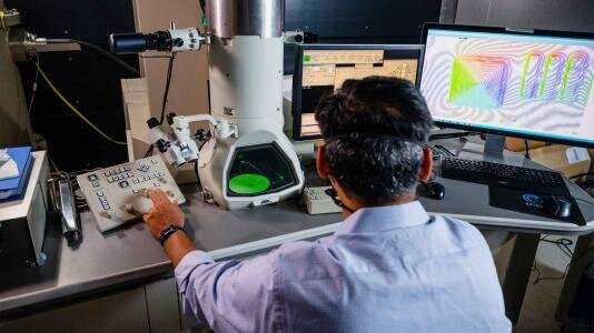
With resolution 1,000 times greater than a light microscope, electron microscopes are exceptionally good at imaging materials and detailing their properties. But like all technologies, they have some limitations.
To overcome these limitations, scientists have traditionally focused on upgrading hardware, which is costly. But researchers at the U.S. Department of Energy’s (DOE) Argonne National Laboratory are showing that advanced software developments can push their performance further.
Argonne researchers have recently uncovered a way to improve the resolution and sensitivity of an electron microscope by using an artificial intelligence (AI) framework in a unique way. Their approach, published in npj Computational Materials, enables scientists to get even more detailed information about materials and the microscope itself, which can further expand its uses.
“Our method is helping improve the resolution of existing instruments so people don’t need to upgrade to new expensive hardware so often,” said Argonne assistant scientist and lead author Tao Zhou.
Challenges with electron microscopy today
Electrons act like waves when they travel, and electron microscopes exploit this knowledge to create images. Images are formed when a material is exposed to a beam of electron waves. Passing through, these waves interact with the material, and this interaction is captured by a detector and measured. These measurements are used to construct a magnified image.
Along with creating magnified images, electron microscopes also capture information about material properties, such as magnetization and electrostatic potential, which is the energy needed to move a charge against an electric field. This information is stored in a property of the electron wave known as phase. Phase describes the location or timing of a point within a wave cycle, such as the point where a wave reaches its peak.
When measurements are taken, information about the phase is seemingly lost. As a result, scientists cannot access information about magnetization or electrostatic potential from the images they acquire.
“Knowing these characteristics is critical to controlling and engineering desired properties in materials for batteries, electronics and other devices. That’s why retrieving phase information is important,” said Argonne material scientist and group leader Charudatta Phatak, a co-author of the paper.
Using an AI framework to retrieve phase information
Retrieving phase information is a decades-old problem. It originated in X-ray imaging and is now shared by other fields, including electron microscopy. To resolve this problem, Phatak, Zhou and Argonne computational scientist and group leader Mathew Cherukara propose leveraging tools built to train deep neural networks, a form of AI.
Neural networks are essentially a series of algorithms designed to mimic the human brain and nervous system. When given a series of inputs and output, these algorithms seek to map out the relationship between the two. But to do this accurately, neural networks have to be trained. That’s where training algorithms come into play.
“Tech companies like Google and Facebook have developed packages of software that are designed to train neural networks. What we’ve essentially done is taken those and applied them to the scientific challenge of phase retrieval,” said Cherukara.
Using these training algorithms, the research team demonstrated a way to recover phase information. But what makes their approach unique is that it also enables scientists to retrieve essential information about their electron microscope.
“Normally when you’re trying to retrieve the phase, you presume you know your microscope parameters perfectly. However, that knowledge might not be accurate,” Zhou pointed out. “With our method, you don’t have to rely on this assumption. Instead, you actually get the conditions of your microscope—that’s something other phase retrieval methods can’t do.”
Their method also improves the resolution and sensitivity of existing equipment. This means that researchers will be able to recover tiny shifts in phase, and in turn, get information about small changes in magnetization and electrostatic potential, all without requiring costly hardware upgrades.
“Just doing a software upgrade we were able to improve the spatial resolution, accuracy and sensitivity of our microscopy,” said Zhou. “The fact that we didn’t need to add any new equipment to leverage these benefits is a huge advantage from an experimentalist’s point of view.”
The paper, titled “Differential programming enabled functional imaging with Lorentz transmission electron microscopy,” was published Sept. 6.