The NexGen 7T MRI scanner marks a breakthrough in brain imaging, providing unprecedented resolution that could transform neuroscience research and the diagnosis of brain disorders. Funded by the BRAIN Initiative, this technology enables detailed imaging of brain circuitry and could lead to significant advancements in understanding mental and neurological disorders.
Higher resolution will allow neuroscientists to more precisely localize and trace brain networks.
An intense international effort to improve the resolution of magnetic resonance imaging (MRI) for studying the human brain has culminated in an ultra-high resolution 7 Tesla scanner that records up to 10 times more detail than current 7T scanners and over 50 times more detail than current 3T scanners, the mainstay of most hospitals.
Enhanced Detail in Brain Imaging
The dramatically improved resolution means that scientists can see functional MRI (
“The NexGen 7T scanner is a new tool that allows us to look at the brain circuitry underlying different diseases of the brain with higher spatial resolution in fMRI, diffusion and structural imaging, and therefore to perform human neuroscience research at higher granularity. This puts UC Berkeley at the forefront of human neuroimaging research,” said David Feinberg, the director of the project to build the scanner, acting professor at the Helen Wills Neuroscience Institute at the 
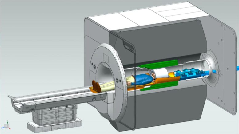
Cross-sectional diagram of the NexGen 7T scanner, showing the new Impulse head-only gradient coil (green) and receiver-transmit coil (white) resting on a movable bed (brown) and connected to an electronic interface (blue) containing nearly a thousand wires (blue) that extend out of the magnet. Credit: Bernhard Gruber, MGH Harvard
A New Era in Neuroscience Research
The improved resolution will help neuroscientists probe the neuronal circuits in different regions of the brain’s neocortex and allow researchers to track signals propagating from one area of the cortex to another as we think and reason, and perhaps discover underlying causes of developmental disorders. This could lead to better ways of diagnosing brain disorders, perhaps by identifying new biomarkers that would allow diagnosis of mental disorders earlier or, more specifically, in order to choose the best therapy.
“Normally, MRI is not fast enough at all to see the direction of the information being passed from one area of the brain to another,” Feinberg said. “The scanner’s higher spatial resolution can identify activity at different depths in the brain’s cortex to indirectly reveal brain circuitry by differentiating activity in different cell layers of the cortex.”

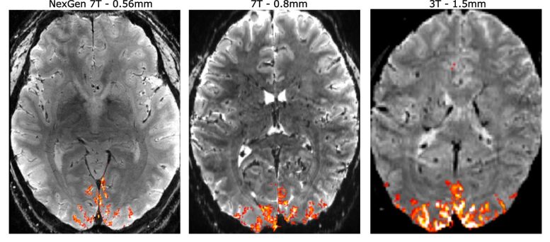
A comparison of human brain scans using the NexGen 7T MRI at higher resolution (left) vs a conventional 7T scanner (middle) and the standard 3T hospital scanner (right). With higher resolution, neuroscientists can more precisely localize signals (orange) in the brain to understand the normal brain circuitry and the changes associated with brain disorders. Credit: David Feinberg and Alex Beckett, UC Berkeley and Advanced MRI Technologies
This is possible because neuroscientists have found in vision brain areas that the superficial and deepest cortex layers (blue arrows in image on right) incorporate “top-down” circuits, that is, they receive information from higher cortical brain areas, whereas the middle cortex involves “bottom-up” circuitry, receiving input to the brain from our senses. Pinpointing the fMRI activity to a specific depth in the cortex lets neuroscientists track the flow of information throughout the brain and cortex.
With the higher spatial resolution, neuroscientists will be able to home in on the activity of something on the order of 850 individual neurons within a single voxel — a 3D pixel — instead of the 600,000 recorded with standard hospital MRIs, said Silvia Bunge, a UC Berkeley professor of psychology who is one of the first to use the NexGen 7T to conduct research on a human brain.
“We were able to look at the layer profile of the prefrontal cortex, and it’s beautiful,” said Bunge, who studies abstract reasoning. “It’s so exciting to have this state-of-the-art, world-class machine.”
Revolutionizing Brain Disorder Research
For William Jagust, a UC Berkeley professor of public health who studies the brain changes associated with 
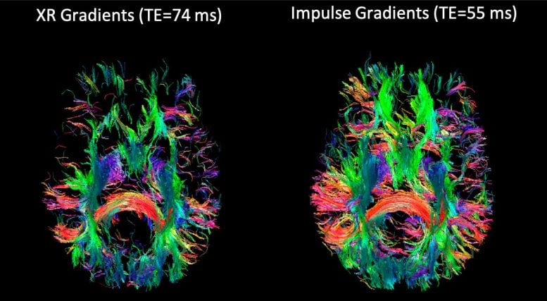
Diffusion MRI imaging — called tractography — of the bundles of axons that neurons send throughout the brain’s white matter, which lies interior to the cortex gray matter at the surface of the brain. The images reveal the communications circuitry extending throughout the white matter from the surrounding cortex. Credit: An T. Vu, UCSF, and David Feinberg, UC Berkeley, and Advanced MRI Technologies


